|
4:00 P.M.
February 28, 2002
Mysteries & Phantoms
Of the Brain
by Myron J. Talbert M.D.
Assembly Room, A. K. Smiley Public Library
Summary
This paper discusses various conditions involving the brain. Based on research by my
father, George A. Talbert. Ph.D. in Berlin, Germany in 1898, in which he used a new
technique of demonstrating the motor areas of the brain in dogs. Cases are described from
two books, Phantoms of the Brain by Ramachandran & Blakeslee and The Man Who Mistook
his Wife for a Hat by Oliver Sacks M.D. I described my own experience with a stroke.
Information about current treatments for mental illness, Alzheimer's disease and stroke
was obtained from the internet National Library of Medicine.
Background of the Author
Myron J. Talbert was born in Omaha, Nebraska. He grew up in Grand
Forks, North Dakota, from age 1 1/2. After college graduation from the University of
North Dakota, attended the first two years of Medical school. Then he transferred to
Temple Univ. School of Medicine ,graduating in 1946. After Internship at Madison General
Hospital, he served 2 years in the army as a 1st Lt. He returned to Madison where he
completed Surgical Residency in 1953. He practiced in Grand Forks, N.D. From 1956.
until retirement in December, 1989, he prcticed general surgery in Redlands, California, ,
serving as Chief of staff of the Redlands Community Hospital twice, and as Chief of staff
at San Bernardino County Hospital. He is a past president of the San Bernardino County
Medical Societyand a past President of the Tri County Surgical Society. He is Board
Certified in General Surgery, and a Fellow of the American College of Surgery. He has
served on several boards in Redlands, including the Redlands Art Assn (president), Red Cross, Salvation Army (president), Redlands Community Music Assn (Redlands Bowl). He
has been a member of the Noon Kiwanis Club since 1956 and the program chairman for 11
years. His wife, Harriet, and he enjoy their three daughters and their families.
Mysteries and Phantoms of the Brain
The subject, mysteries and phantoms of
the brain, is of interest to me for various reasons. First of all my father, George A.
Talbert Ph.D. had done some early research in brain localization in 1898 with Dr Hermann
Monk in Berlin, Germany.
The second reason is obviously related
to my own experience with a cerebral vascular accident on September 1st, 2000,
and yet another reason is I have always wondered what makes Harriet’s brain tick. The
latter may never be answered but there is something different about a person who can play
a piano by reading two lines and three measures ahead. In the case of the organ she must
read three lines, one for each hand and one for the feet. I think there most be a special
modem implanted in her brain to accomplish that.
Although, the areas of localization of
motor function, were roughly known, my father devised a different method of demonstrating
these areas by implanting ivory plugs with electrodes in the skulls of dogs then
stimulating those areas after the dog had recovered from the anesthesia.
Fritsch
and Hitsig reported their work in 1871 by electrical stimulation of the areas of motor
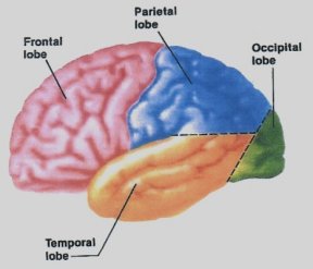
function located anterior to the fissure of Rolando in the parietal lobes of the brain..
Richard Ewald from Strasburg had reported similar findings. My father’s work modified Ewald’s technique and was reported in
the Philadelphia Medical Journal November 25, 1899.
In the 1940s and 50s a brilliant
Canadian neurosurgeon, Wilder Penfield, mapped out sensory areas in the brain on humans in
the course of neurological operations done under local anesthesia. He was able to do this
because, although the brain is entirely nervous tissue, it is not sensitive to pain. He
was then able to map out a “homunculus” or little man that one could imagine,
lies over the brain and represents the sensory areas (see illustration #1).
He
is upside down for the most part and his feet are tucked in the bottom of the medial
surface of the parietal lobe near the top whereas the face is down near the bottom of the
outer surface. The face and hands occupy a relatively large proportion of the map, no
doubt, due to their greater sensitivity than that of the trunk.
A
strange thing is that the face area is below the hand instead of being near the neck and,
Oddly enough, the genitals are located near the feet. I will discuss this more later
.It is interesting that in my fathers
work, he described, in addition to leg movements, there was often movement of the tongue
and facial expression in the form of barring the teeth when the brain was stimulated with
an electric current. He, of course, didn’t have the information of the later work of
Penfield about how near the face is to the shoulder area.
My personal experience with the stroke
was difficult for me to diagnose. I knew something was wrong because while playing golf I
had rather severe low back pain and weakness but I thought, since I had experienced
previous back pain, that was the problem. I called my physician who then saw me in his
office.
I was able to walk in and. I had no
problem with my left arm at that time so he also thought it was my back that was causing
the problem. So I went home. It was then that I developed a foot drop causing me to fall
down, fortunately without injury. My left arm then became weak so that clinched the
diagnosis and, therefore, I went directly to the hospital emergency room where the ER
doctor did an excellent physical, ordered a Brain CT scan, an MRI, an ultra sound of my
carotid arteries and an EKG.
They
were able to discern that I had an intravascular clot that involved only a small, but
critical, artery, causing a left hemi-paralysis with no evidence of hemorrhage.
The
carotid arteries are often the source of clots that break off and lodge in arteries in the
brain. If these are involved then the neck should be opened and the lining of the involved
carotid artery cleaned out. That is why an ultrasound study of the neck is important.
There is a new treatment that can be
used in cases such as mine which involves injection of a medication that dissolves the
clot called ATP.. The results have been good as far as recovery is concerned, however it
can be used only within a three-hour window from the time of the first symptoms otherwise
there is a danger of hemorrhage
Since
my symptoms began at about 2PM and it was 8PM when I was admitted.. The treatment had to
be rehabilitation and the taking of aspirin with a drug called plavix to prevent further
clotting. This was started immediately.
The
clinical evaluation was interesting as they kept asking me what day, month, and year it
was, who is president and where am I. They also wanted to know what 100 minus 7 was. I was
sorry that I didn’t study for the exam. They sounded like foolish questions but
actually there are areas in the brain that involve memory, both recent and long term as
well as mathematic skills.
When strokes occur they result from
blockage of an artery either from a clot that forms in an artery of the brain due to a
localized arteriosclerotic plaque as occurred in my case, the breaking off of a clot
either from one of the carotid arteries with embolus to an artery in the brain or from a clot which may form on the inner wall of
the heart after a heart attack with breaking off of a portion of that clot. Otherwise it
may be due to a rupture of an artery in the brain causing a hemorrhage.
Where
the brain is damaged is the key to what disability the patient will have. Sometimes it
affects the motor areas alone or if it is on the left side it may affect the speech area
as well causing aphasia
. Some patients are unable to
comprehend what is said they. often are
not recognized as having aphasia because they do recognize the tone and inflection as well
as the facial expression and body language and thus get the understanding of what was said
even though they don’t understand the words. This is not unlike a person addressing a
dog. He doesn’t understand the words but does get the feeling from the inflection.
Some know what they want to say but cannot remember the words. This form is most
frustrating. The worst is the global form which knocks out everything making communication
impossible.even writing
The treatment involves a speech
pathologist early in the course of the illness .
. They are doing some interesting
studies in Bethesda, Maryland using PET scans and radioactive substances that show local
brain function, in addition drugs are being tested to benefit these patients
When
there is paralysis, there is a cross over so that lesions on the right cause paralysis on
the left and on the left the converse. The brain is also subject to traumatic injuries or
deprivation of oxygen due to shock or carbon monoxide poisoning.
.The brain does not tolerate oxygen
deprivation for very long so treatment designed to relieve the vascular obstruction must
be started as soon as possible. That is why the 3-hour limit in the case of intravascular
clots.
A very recent study indicated that when
an artery is obstructed a substance called glutamate is released which poisons the
surrounding cells and the stroke therefore extends. Eighty five percent of strokes show
this extension in the following 12 weeks
An
experimental drug called citicoline appears to prevent some of this effect. When the lower
dose is given of 500 mg the extension was 34% and when 2000 mg was given only 2%
experienced extension. Unfortunately the drug is not available as yet and the research is
lacking in funds to make it available. It takes a long time to gain approval for new
drugs.
There are other treatments that are
still in the early stages of research. One is that of hypothermia. It is known that people
who drown in cold water seem to tolerate the lack of oxygen to the brain for longer
periods of time so this might have some promise. The other is the use of trans-cranial
magnetic stimulation. This involves the use of a magnetic loop applied over the damaged
area of the skull. This sounds a little far fetched however.
Now I know I have interruption of the
motor pathways due to my stroke, but what I don’t know is; is this a permanent thing
or can one develop new pathways? This is called plasticising. It does happen in children
according to a report on television which showed children with severe epilepsy who had
undergone a complete lobectomy on one side. Surprisingly they were able to walk but had
some apparent weakness of the contralateral arm. They were able to speak with moderate
difficulty, and seemed to not have any impairment in their intelligence. The developing
brain seems to adjust better than the more mature one
An unanswered, but frequently occurring
phenomena, and one that I also experienced, is when I would yawn the paralyzed muscles
would contract, causing my left arm and leg to flex. This seems to be something that
results from using another pathway from the brain temporally. It should be noted that this is a spastic
paralysis rather than a flaccid one, because the upper motor neuron is involved. This means that one has to overcome the tightness
of the muscles when learning to move the extremities.
Because of my experiences my interest
was piqued to study other aspects of brain function. This led me to research done by Dr. Ramachandran, a neuro-psychiatrist, at
U.C. San Diego. He and Sandra Blakeslee had a
special interest in phantom pains in amputated extremities.
Following the civil war there was a
large number of amputees. This had resulted from the fact that because of the high
infection rate they had found that they saved more lives by amputating. Many of the
patients experienced this distressing phantom pain following the amputation.
The
treatment had consisted of reamputation or removal of the neuroma that they thought was
the cause of the pain. but the result was that many had recurrence of the pain.
Dr
Ramachandran studied an arm amputee who had phantom pain in the amputated arm. He
stimulated the face and upper arm and found that he could map out the hand on both the
face and on the upper arm. by lightly touching those areas with a q tip and the patient
would feel it on face as well as on the hand even though it was no longer there. He went a
step further by wetting the q tip with warm water and the patient volunteered that he felt
the warm water run down his phantom hand
See illustration 2. These areas fit the
pattern of the homunculus man that Dr Penfield described. The answer lies in the peculiar
mapping of the body part with the face lying right beside the hand. Furthermore the hand
area is flanked below by the face area and above by the shoulder area. Dr Ramachandran
verified his findings on the same patient using magnetoencephalography. This is an MRI
that shows brain activity. This explains why reamputation or excision of the neuroma
rarely is effective.
Some patients with phantom pain also
say they feel that they can move the non-existent extremity. What then happens with the
patient born without arms? Dr Ramachandran had the opportunity to study such a patient.
She was 25 years old and was born without arms but claimed she used her arms to
gesticulate and even point at things. Her arms were only small projections where the arms
should be. She wore prostheses but felt they should be made shorter than normal. This
suggests that she had some neural circuitry set down, at least partially, by the genes. So
is it then nature rather than nurture that controls these feelings?
Another patient, age 55 had his forearm
amputated for cancer. In addition to the pain he experienced the sensation of the hand
going into spasm with nails digging into the palm. Dr Ramachandran devised a box with a
mirror in it so that when he put his normal hand in it the patient could see the mirror
image representing the amputated hand. He would have the patient open his normal hand and
the spasm was relieved. He tried this technique in a dozen patients with similar problems
and it worked in half of them.
It is hard to know if this is the
placebo effect or not as many of these patients are susceptible to placebos, however, 50%
is higher than expected for placebo effect.
He also had another patient, who had a
below the knee amputation. This patient also verified Penfield’s little man map when
she volunteered the information that she experienced a sensation in her foot during sexual
intercourse. Remember that the Penfield map shows the genital area is adjacent to the feet
People respond differently to strokes,
some have severe headaches, especially if hemorrhage is involved and many are obtunded.
Others, such as myself, have no head symptoms.
Patients with left sided strokes tend
to be more anxious and depressed by their condition while those with right sided lesions
tend to be indifferent to their problem. It is a known fact that the right hemisphere
tends to be more emotionally volatile than the left. This is thought to be the difference
between men and women as well. Women tend to use their right brain and are more interested
in the home life of an athlete than they are with his batting average. One thing I have experienced is I am a little
more emotional. I sometimes cry when the Rotolo Chevrolet add comes on.
Patients react differently to their
disability. There are some patients that deny that they have any paralysis even though
they cannot move their extremities on one side of their body. They may go so far as to say
that the arm in their bed belongs to someone else, yet they are oriented in other ways.
One patient was found on the floor repeatedly. The reason, it turned out, was due to the
fact he kept trying to throw the strange arm out of the bed and, of course, the body went
with it.
. In a similar manner some patients
have what is known as a hemi neglect syndrome. When tested they are not blind in one
hemisphere of vision as they may see motion in a finger in that hemisphere but they
disregard the area. This syndrome occurs when the stroke involves the right parietal
lobe... One female patient would make up
only half her face and also would eat only half of the food on her plate. .
Oliver Sacks M.D., Professor of
psychiatry at Albert Einstein School of Medicine in New York, has written an interesting
book called “The Man Who Mistook His Wife For A Hat” in which he describes
different effects of strokes.
In
that particular case the patient was unable to identify objects and when he sought to put
his hat on he grasped his wife’s head thinking that it was his hat. He was unable to
identify objects such as a glove. He could
describe it, but it was difficult for him to figure out how it is used. In addition, he was unable to recognize family
faces.
Some patients develop epileptic
seizures. One 88 year old awoke following a vivid dream of her youth in Ireland in which
she heard nostalgic songs that she had loved. Oddly enough when she awoke the music
persisted. This persisted for several weeks. An EEG was done which showed a definite
indication of temporal lobe seizures and a brain scan confirmed the presence of an infarct
in the temporal lobe. This patient enjoyed the experience so much that she declined
treatment. A second similar case had similar findings on brain scan but the music was so
disturbing that she begged for treatment. She was given anticonvulsant medication, which
controlled the condition.
Neither
of these patients had any convulsions which is characteristic of most epileptic patients.
Dr
Sacks goes on to mention two other famous people with similar sensations. The first was
Shostakovich, described in an article in the New York Times under the title of “Did
Shostakovich Have A Secret?” In the article he was said to have a shell fragment in
the temporal lobe. It was said that he could hear music when he tilted his head. He seemed
to enjoy the sensation and did not want to have it removed.. This was a case of trauma, of
course, rather than a stroke.
Dostoevsky was said to have psychical
seizures or elaborate mental states before a seizure. He said “you healthy people,
can’t imagine the happiness which we epileptics feel during the seconds before our
fit---I don’t know if this felicity lasts for seconds, hours, or months, but believe
me I would not exchange it for all the joys that life may bring “(T. Alajouanine,
1963)
Penfield found in his research that
when the temporal lobe on the right is stimulated electrically the patient may hear music
which would explain these findings
A Physician by the name of Hughlings
Jackson in the 1880s first described focal convulsions that arise from localized areas in
the brain. These cases might in some way fall into that category. These are known as
Jacksonian seizures. During these seizures the patient doesn’t lose consciousness.
How the brain handles vision is
remarkable in as much as what we see is an inverted image and the brain must correct it
but perception involves more than replication of an image in the brain. Take the Necker
cube. As you look at it, it is as though you are looking down on it then spontaneously it
will change to appear to be above you. Thus there is an element of judgment involved. See
illustration #3 Another classic illusion is that of the two circles that are of the same
size. One, however, is surrounded by large circles and the other by small ones. For most
people the one surrounded by the large circles looks smaller than the one surrounded by
the small circles. Illustration #4 This illustrates that perception is relative.
We all have blind spots in each eye,
but we never notice them because the other eye fills in the spot. If you put two spots on
a paper cover one eye, gaze at one spot then bring the paper slowly in toward the face,
the lateral spot disappears. This occurs because the image falls on the optic nerve of
that eye where there are no receptors illustration #5.. An interesting thing occurs if one
draws a line through the lateral dot but off set the lower part the mind will correct it
and the line appears to extend straight through. Illustration #6 This correction does not
occur if the offset is vertical because our eyes are beside each other rather than one
above the other.
The visual pathways are extremely
complicated, but basically if you look straight ahead the entire world on your left is
mapped into your right visual cortex.. An interesting thing may happen .in patients with a
visual field blindness they are able sometimes to reach for and grasp an object in the
blind area. or to turn an object such as a letter to fit it into a slot yet they cannot
actually see it consciously.. Some call this “blind sight” This is thought to be
due to the presence of two pathways, one old, phylogenetic one and a newer pathway. They
are known as the “how” and the “what” pathways. Somehow these people
use one of them.
James Thurber was blinded in one eye
when he was six years old and by the age of thirty-five his other eye went blind. But his
visual field, instead of being black was filled with visual hallucinations. His blindness
was filled with pixie dust and brightness. He had what is known as Charles Bonet Syndrome.
Apparently this condition is not uncommon and it occurs in people with glaucoma, macular
degeneration and diabetic retinopathy. The hallucinations may involve, as they did with
Thurber, seeing a gay old lady with a gray parasol walk through the side of a truck Some
such patients don’t report it for fear of being considered crazy. Many of these
patients can cause the hallucination to disappear when they close their eyes. It is
thought that Thurber got his ideas for cartoons from his visual hallucinations’
. Patients with migraine will have a
prodromal scotoma or blind spot. For example if they are looking at a grand father clock
standing against a wall with wall paper on it the clock will disappear but there will not
be a black area where the clock is but the mind will fill the blind spot with the rest of
the wall paper. Thus vision involves some interpretation by the brain.
We have talked about patients with
damaged areas of the brain, however, there is another group with excesses.
In 1885 Dr Gilles de la Tourette
described a syndrome that bears his name. This is characterized by excess of nervous
energy with tics, jerks, grimaces , curses and loud explosive noises. In the milder forms
of the disease many patients are able to control it with a drug called Haldol.
Dr Sacks described a patient he
called witty ticcy Ray who suffered from Tourettes. He was highly intelligent, a
college graduate and was married. He was
quite popular due to his quick wit and because of the characteristic excessive energy of
this syndrome, he was an excellent drummer with terrific licks. Because of his excellent timing, he was also a
very fine ping-pong player. He had this condition since the age of four and was
twenty-four when he was first seen by Dr Sacks. While
the energy inherent with the syndrome was a great asset to his drumming and ping pong, the
ticks and swearing caused him to lose several jobs. And
therefore he sought help.
The disease is related to an excess of
dopamine in the brain where as Parkinson’s has a lack of it. So in some respects the
two are some how related .
Dr Sacks put the patient on haldol and, although he was given a low, dose he
returned with a black eye and stated “so much for your frigging Haldol.” What had happened was he had lived with the
accelerated reactions since the age of four but, when his reactions were slowed down by
the drug, he was unable to negotiate a revolving door. His tics were controlled, however,
and after three months of psychotherapy another attempt was made with the Haldol. He then
found a happy medium by taking the drug during the working week and stopping it on the
weekend. “
A paper on this subject would be remiss
if the subject of Alzheimer’s disease was not addressed. There are about 4 million
people with the condition. Although it can occur in younger people especially with
familial genes it is most common between the ages 65 to 74 at which time 3% of the
population will have it. This jumps to 50% over age 85. If, however, you reach 90 you are
not likely to be afflicted.
I always reassured my patients that I
would diagnose their case if it takes an autopsy to do it. Actually that has been true of
this disease until recently. Now, making use of MRI and PET scans, many centers have been
able to diagnose it in 90% of the cases.
The disease is due to the deposition of
an amyloid like material at the neural connections in the form of plaques and tangles,
which causes loss of memory and other cognitive skills.
The first symptoms usually are those of
forgetfulness, which progresses to inability to identify objects and to recognize faces,
even family members. They may be confused and will often wander off and get lost. So they
require close supervision. Sometimes they may
become belligerent and need medication to calm them down.
The disease in itself, doesn’t
cause death as a rule, but problems with swallowing of food causes aspiration that results
in pneumonia that frequently is the cause of death.
There doesn’t seem to be a
standard treatment for Alzheimer’s disease, but there are three or four drugs that
have been tried in an effort to prevent the deposit of the amyloid substance. These and
newer drugs need further investigation.
There is one other group of diseases
that affect the brain and that is the psychosis such as manic depressive or bi polar
disease. This condition may occur at any time in life, but is commonly seen at
adolescence. It is characterized by severe mood swings from deep depression and ideas of
suicide to hyper function and activity. Drug abuse frequently accompanies the condition. Lithium is commonly used to treat the condition
together with psychotherapy.
. There are 1.3 million Americans with
this illness both male and female many of whom, with therapy, are able to function quite
well.. It is commonly seen in families, so researchers are attempting to find the gene
with the hope to be able to alter that gene to effectively control it. Here is where gene
engineering may be a Godsend. I hope we don’t lose sight of the benefits of this
procedure thinking that we are second guessing our maker. Perhaps the study of stem cells will also be helpful in diagnosing and
treating this tragic illness.
Shortly after I retired I was asked by
the Attorney General to evaluate a young man for disability who had been stabbed by a
fellow inmate in prison. He developed a severe wound infection called necrotising
fasciitis after the surgery that had left him with no abdominal muscles, so the disability
was severe.
In
tracing the story he was convicted of armed robbery at the age of 17. He had been sent to
a juvenile facility first, but could not be controlled there so he was sent to an adult
prison. Actually, he had served his time when he was stabbed as he was leaving the prison.
He was studied after his near death experience and found to have severe bipolar disease.
One wonders how his life might have been changed if the diagnosis had been made before he
got into trouble.
Another mental illness, schizophrenia,
appears to be due to a defect in the prefrontal lobe of the brain. Studies using a PET
scan indicate that there is decreased function in this area when the patient is given
working memory tests and abstract reasoning. This defect causes increase in release of
dopamine which is the substance that causes the delusions and hallucinations. The drugs
that are used quell the excess dopamine, Haldol, has been used, but newer drugs such as
Clozaril have been more effective. Recent reports indicate, however, severe side effects
such as inflammation of the heart can occur with this drug. It occurs in less than 1% of
the cases but the consequences could be fatal.
Ziprasidone is one of the latest drugs
but is not released as yet. It blocks the dopamine release, but side effects in large
numbers of patients are still to be studied. One
strong recommendation is that the best results are obtained when treatment is given early
in the course of the disease combined with psychotherapy.
The PET (positron emissions tomography)
together with radioactive form of oxygen are remarkable studies of the brain that light up
when a specific area of the brain is used. Thus more exact information is obtained which
never before was available.
Well, what have I learned from all
this? I know I’m going to try to squeeze out some more function out of this lame
brain of mine and there is something special in Harriet’s head, and maybe now I
understand why she turns to the left at the brown house on the corner rather than turning
west on Olive and maybe why she turns the heat off rather than setting the thermostat
down, The only answer there, I guess, is she uses her right brain when I am using my left.
|